Navigation auf uzh.ch
Navigation auf uzh.ch

When PhD student Gabriele Gut does an experiment, he’s not just looking at a few cells under a microscope. He’s looking at hundreds of thousands at once. And instead of investigating the cells with a light or electron microscope, he’s using a Yokogawa machine, an automated microscope as big as a medium-sized box. The machine’s optics allow every single cell marked with fluorescing chemicals to be scanned with light of different frequencies.
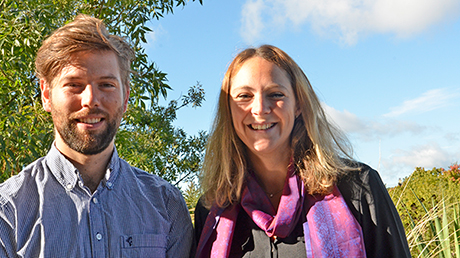
The gigabytes of microscopic data output by the machine then go to the nearest supercomputer for evaluation. This is where Cycler comes in, a program developed by Gut and his thesis supervisor Prisca Liberali. A day later Gut can admire the latest curves conjured up by his program. They show how crucial biological properties change in the course of the cell cycle. “Computer-based image analysis gives unprecedented insights into what happens in cells,” says Gabriele Gut.
Simple but not oversimplifying
Welcome to the world of quantitative biology, an approach quietly finding its way into life sciences labs to enable new, even revolutionary, ways of looking at biological processes. “Without quantitative biology we’re not going to get much further in molecular and cellular biology,” explains Prisca Liberali. Take Cycler, which Gut and his mentor at Lucas Pelkman’s lab have developed in collaboration with scientists at Columbia University in New York: It enables variations in biological properties ascertained from snapshots of hundreds of thousands of cells to be used to represent the entire cell cycle of the population. “It’s a simple method, but it doesn’t oversimplify things,” says Liberali. The team has just been able to publish its initial studies with the innovative program in the prestigious specialist journal Nature Methods.
Cycler can analyze cells on the basis of snapshots to find out how the quantity of an important regulating protein changes in the course of the cell cycle. Only then does it becoming clear, for example, that a regulating protein is only synthesized in large amounts before the cell divides. The activities of genes analyzed using this method also display variations over the cell cycle.
Big data in biology
When asked why this is important, Prisca Liberali answers: “To better understand how cells function we have to know about the dynamics of individual cellular processes.” For example quantitative screening of infection with a virus – the SV40 virus – has shown that infectivity depends on the size of the cell nucleus. This completely unexpected insight has only been possible thanks to this mass screening.
It’s also conceivable that findings of this sort might throw new light on biological processes, as well as explaining why certain results have not been reproducible in the past. To continue our example: A virologist conducting infection experiments with cells that by chance have small nuclei will draw the wrong conclusions.
This new approach in biology will increase the importance of exact scientific disciplines such as mathematics, physics, and programming. “We’re still biologists, not mathematicians,” say Prisca Liberali and Gabriele Gut, “but collaboration across disciplines is essential.” For example Michelle Tadmor, who helped with programming the algorithms and co-authored the Nature paper, is a mathematician. “It’s important for us to understand each other’s specialist languages,” say the two biologists. For them it’s clear that for researchers at the forefront of biology, there’s simply no way around an in-depth knowledge of statistics and programming.
.